
Файл:Prognathodon tissue.jpg
Размер этого предпросмотра: 789 × 600 пкс. Другое разрешение: 1632 × 1241 пкс.
Исходный файл (1632 × 1241 пкс, размер файла: 263 КБ, MIME-тип: image/jpeg)
Описание
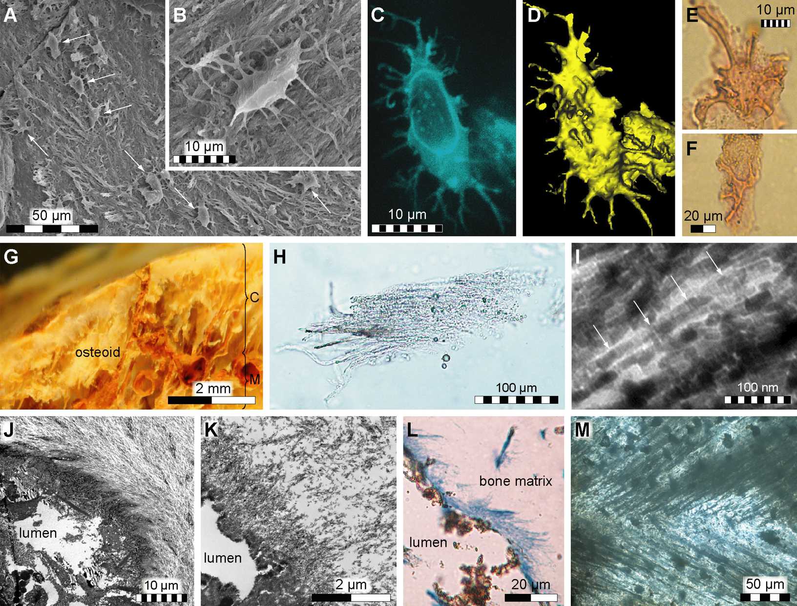
| Описание | Fibrous tissues and microstructures recovered from IRSNB 1624.
|
|---|---|
| Источник | http://www.plosone.org/article/info%3Adoi%2F10.1371%2Fjournal.pone.0019445 |
| Время создания | 2011 |
| Автор или правообладатель | Johan Lindgren1*, Per Uvdal2,3*, Anders Engdahl2, Andrew H. Lee4, Carl Alwmark1, Karl-Erik Bergquist5, Einar Nilsson5, Peter Ekström6, Magnus Rasmussen7, Desirée A. Douglas6¤, Michael J. Polcyn8, Louis L. Jacobs8 — Лицензия: CC BY 2.5 (Creative Commons Attribution 2.5) https://creativecommons.org/licenses/by/2.5 |
| Другие версии файла | — |
Источник файла — сайт Wikimedia Commons, куда он был загружен под одной из свободных лицензий ( https://commons.wikimedia.org/wiki/File:Prognathodon_tissue.jpg ). Авторов, работавших над этим файлом см. в истории файла: https://commons.wikimedia.org/w/index.php?title=File:Prognathodon_tissue.jpg&action=history
В общем случае в статьях энциклопедии Руниверсалис файлы используются в соответствии со статьёй 1274 Гражданского кодекса Российской Федерации.
История файла
Нажмите на дату/время, чтобы увидеть версию файла от того времени.
| Дата/время | Миниатюра | Размеры | Участник | Примечание | |
|---|---|---|---|---|---|
| текущий | 03:49, 23 ноября 2023 | | 1632 × 1241 (263 КБ) | Я, робот (обсуждение | вклад) | == Описание == {{Изображение | описание = <p>Fibrous tissues and microstructures recovered from IRSNB 1624. </p> <ul><li>(A) Secondary electron micrograph of acid etched cortical bone showing fibrous tissues and what appears to be part of the osteocyte-lacunocanalicular system (osteocyte-like entities at arrows).</li> <li>(B) Osteocyte-like structure in lacuna within fibrous tissues.</li> <li>(C) Isolated osteocyte-like form visualized with fluorescent dye.</li> <li>(D) Topographic image of... |
Вы не можете перезаписать этот файл.
Использование файла
Следующая страница использует этот файл: