
Файл:Hsv encephalitis.jpg
Hsv_encephalitis.jpg (480 × 512 пкс, размер файла: 88 КБ, MIME-тип: image/jpeg)
Описание
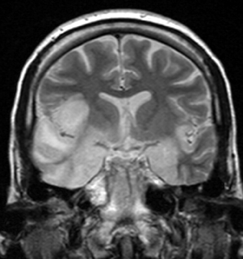
| Описание | "This 33 year-old female patient presented with agitation, confusion, mutism, and fever. This coronal T2-weighted MR image shows high signal in the temporal lobes including hippocampal formations and parahippogampal gyrae, insulae, and right inferior frontal gyrus. The right gyrus rectus and the columns of the fornices were also involved (not shown). There was no associated haemorrhage or enhancement. There was moderate mass-effect. Lumbar puncture was performed - herpes simplex virus DNA was not shown on polymerase chain reaction. A brain biopsy was performed and the histology was consistent with encephalitis. PCR was repeated on the biopsy specimen and was positive for HSV type I." (dr Dawes) |
|---|---|
| Источник | http://www.radpod.org/2007/03/24/herpes-simplex-encephalitis/ |
| Время создания | 2008 |
| Автор или правообладатель | dr Laughlin Dawes — Лицензия: CC BY 3.0 (Creative Commons Attribution 3.0) https://creativecommons.org/licenses/by/3.0 |
| Другие версии файла | — |
Источник файла — сайт Wikimedia Commons, куда он был загружен под одной из свободных лицензий ( https://commons.wikimedia.org/wiki/File:Hsv_encephalitis.jpg ). Авторов, работавших над этим файлом см. в истории файла: https://commons.wikimedia.org/w/index.php?title=File:Hsv_encephalitis.jpg&action=history
В общем случае в статьях энциклопедии Руниверсалис файлы используются в соответствии со статьёй 1274 Гражданского кодекса Российской Федерации.
История файла
Нажмите на дату/время, чтобы увидеть версию файла от того времени.
| Дата/время | Миниатюра | Размеры | Участник | Примечание | |
|---|---|---|---|---|---|
| текущий | 01:46, 24 октября 2023 | | 480 × 512 (88 КБ) | Я, робот (обсуждение | вклад) | == Описание == {{Изображение | описание = <p>"This 33 year-old female patient presented with agitation, confusion, mutism, and fever. This coronal T2-weighted MR image shows high signal in the temporal lobes including hippocampal formations and parahippogampal gyrae, insulae, and right inferior frontal gyrus. The right gyrus rectus and the columns of the fornices were also involved (not shown). There was no associated haemorrhage or enhancement. There was moderate mass-effect. </p> Lumbar pu... |
Вы не можете перезаписать этот файл.
Использование файла
Следующий файл является дубликатом этого файла (подробности):
- Файл:Hsv encephalitis.jpg на общем хранилище
Следующая страница использует этот файл: