
Файл:Doublecortin expression.png
Исходный файл (793 × 1224 пкс, размер файла: 1,4 МБ, MIME-тип: image/png)
Описание
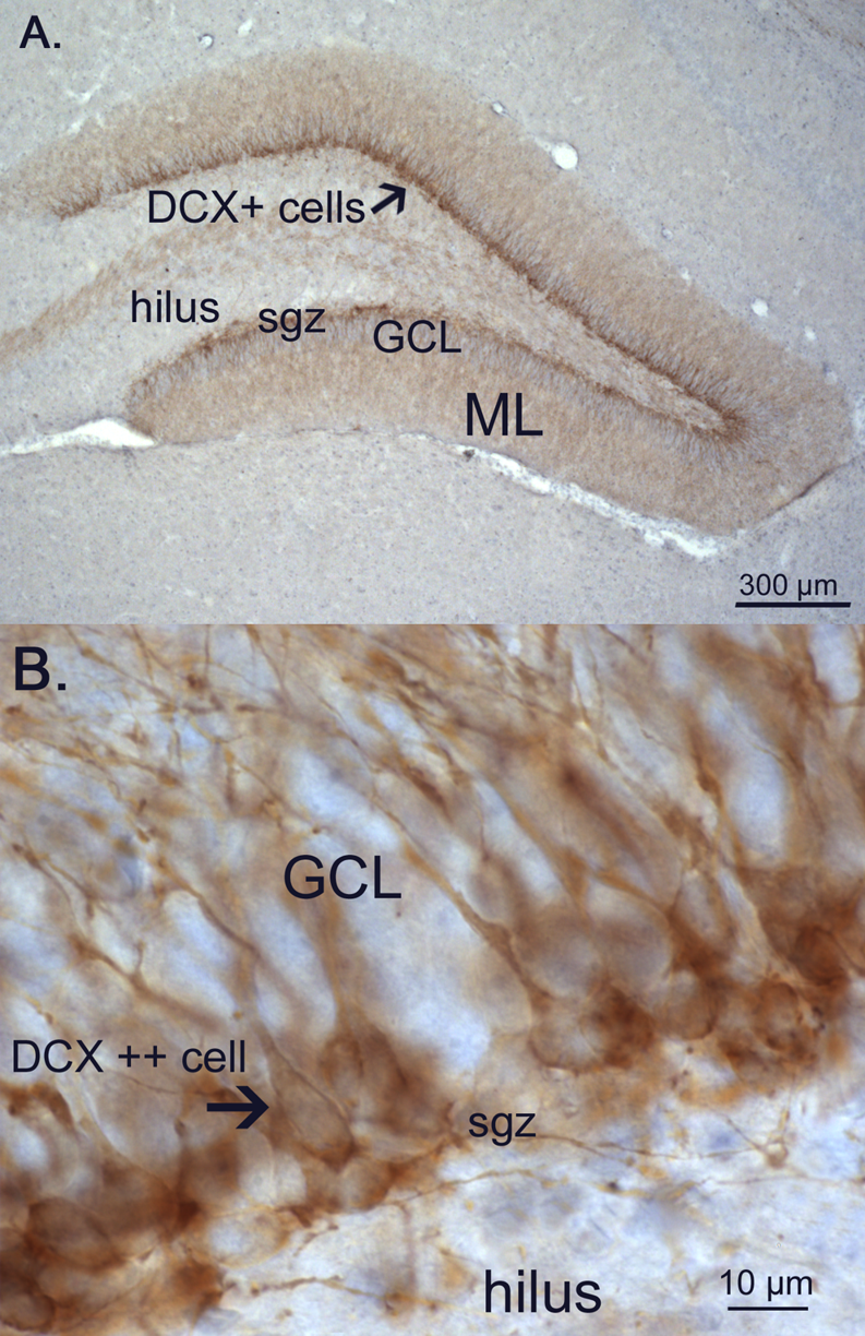
| Описание | Figure 6. Doublecortin (DCX) -positive neurons.
A. Photo of the dentate gyrus of a 21 day old CONS male showing extensive immunostaining of DCX in the subgranular zone (sgz) and the first third of the granular cell layer (GCL) with dendrites extending through the granular cell layer (GCL) into the molecular layer (ML). B. High power photomicrograph showing details of the DCX+ cell bodies located in the SGZ and GCL, with extending dendrites in the |
|---|---|
| Источник | Opposite Effects of Early Maternal Deprivation on Neurogenesis in Male versus Female Rats |
| Время создания | 2009 |
| Автор или правообладатель | Charlotte A. Oomen, Carlos E. N. Girardi, Rudy Cahyadi, Eva C. Verbeek, Harm Krugers, Marian Joëls, Paul J. Lucassen — Лицензия: CC BY 2.5 (Creative Commons Attribution 2.5) https://creativecommons.org/licenses/by/2.5 |
| Другие версии файла | — |
Источник файла — сайт Wikimedia Commons, куда он был загружен под одной из свободных лицензий ( https://commons.wikimedia.org/wiki/File:Doublecortin_expression.png ). Авторов, работавших над этим файлом см. в истории файла: https://commons.wikimedia.org/w/index.php?title=File:Doublecortin_expression.png&action=history
В общем случае в статьях энциклопедии Руниверсалис файлы используются в соответствии со статьёй 1274 Гражданского кодекса Российской Федерации.
История файла
Нажмите на дату/время, чтобы увидеть версию файла от того времени.
| Дата/время | Миниатюра | Размеры | Участник | Примечание | |
|---|---|---|---|---|---|
| текущий | 00:59, 9 октября 2023 | | 793 × 1224 (1,4 МБ) | Я, робот (обсуждение | вклад) | == Описание == {{Изображение | описание = Figure 6. Doublecortin (DCX) -positive neurons. A. Photo of the dentate gyrus of a 21 day old CONS male showing extensive immunostaining of DCX in the subgranular zone (sgz) and the first third of the granular cell layer (GCL) with dendrites extending through the granular cell layer (GCL) into the molecular layer (ML). B. High power photomicrograph showing details of the DCX+ cell bodies located in the SGZ and GCL, with extending dendrites in the | и... |
Вы не можете перезаписать этот файл.
Использование файла
Следующая страница использует этот файл: